HumaDerm® Native SCREEN, Native Human Collagen
HumaDerm Native SCREEN
Human Collagen
SKU: CSP
HumaDerm Native SCREEN are tissue culture plates coated with native human-derived collagen solutions. HumaDerm Native SCREEN is ideal for many applications including coating tissue culture surfaces to support cell attachment and growth.
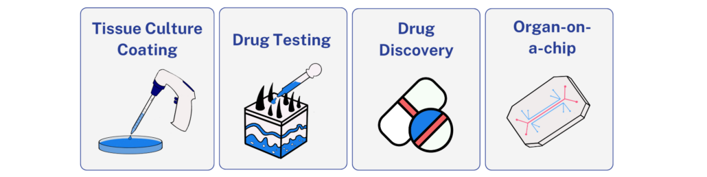
Applications:


")
Background
Product Description
HumaDerm Native SCREEN is a native human collagen screening multi-well plate providing 6 test conditions. HumaDerm Native SCREEN is coated with native human collagen, types I, II, I/III, IV, and V, to help determine which collagen type is best suited for your cell research needs. Each well is designed to deliver precise and independent testing conditions, ensuring the reliability of your data.
Various types of cell surface receptors present on diverse types of mammalian cells recognize the collagen triple helix structure and facilitate cell attachment to collagen materials, including films and scaffolds. The most characterized cell receptors are the integrins α1β1 and α2β1 [2]. Many cell types express both forms of integrin, including mesenchymal stem cells (MSCs), fibroblasts, endothelial cells, chondrocytes, osteoblasts, and lymphocytes [3-6]. Smooth muscle cells interact with collagen via α1β1, and epithelial cells attach via α2β1 [7,8]. There are several types of collagens identified to date, and all are composed of molecules containing three polypeptide chains arranged in a triple helical conformation [1]. The types of collagens differ slightly in the primary amino acid sequence of their polypeptide chains.
Type I collagen is the most abundant type of collagen in the human body, as a major structural matrix protein in skin and many other tissues (bone, tendon, and fibrous connective tissues) [9]. Type I collagen is a heterotrimer composed of one α2(I) chain and two α1(I) chains [3].
Type II collagen is predominantly found in cartilage, the connective tissue that provides structural support and cushioning to joints. It is a critical component of the extracellular matrix in cartilage and plays a significant role in maintaining its structural integrity and function [16]. Type II collagen is a homotrimer composed of three identical α1(II) chains, forming a triple helix structure [17].
Type III collagen is known to be associated with Type I collagen. Collagen type I builds a scaffold with thick fibers that have a low turnover rate. The maturation of collagen type I, however, depends on collagen type III, which produces thin, less durable fibers with a high turnover [10]. Type III collagen conjugated with Type I collagen presents both one α2(I) chain, two α1(I) chains, and three α1(III) chains [11].
Type IV collagen occupies a distinct role within the realm of connective tissues. It is primarily associated with basement membranes, where it plays a pivotal role in mechanical stability and regulates critical functions including cell growth, differentiation, morphogenesis, homeostasis, and regeneration [12]. This collagen type is vital for various physiological processes, including filtration in the kidneys, scaffolding in tissues, and the formation of blood vessel walls. Type IV collagen can exist in at least three hetero-trimeric triple helical forms made up of three α chains [13,14].
Type V collagen is considered a minor component of the fibril-forming collagen family (Type I, II, III, and V) and is primarily found in connective tissues [14]. It interacts with matrix collagens and structural proteins, conferring structural integrity to tissue scaffolds. Type V collagen is broadly expressed as a two α1(V) chains and one α2(V) chain heterotrimer but also as a α1(V), α2(V), and α3(V) heterotrimer in pancreatic islets, adipose tissue, and skeletal muscle [15].
HumaDerm Native SCREEN reduces labor time, consumable costs, and accelerates therapy delivery without affecting the quality of cells.
Tissue Source
The HumaDerm used in HumaDerm Native SCREEN is isolated from human tissue sourced strictly from American Association of Tissue Banks (AATB) accredited and FDA registered tissue banks and organ procurement organizations (OPOs). Humabiologics strives to meet research needs by providing high quality biomaterials obtained from tissue partners who comply with requirements for transplantable human tissues under 21 CFR 1271 of the FDA.
Precautions and Disclaimer
The HumaDerm used in HumaDerm Native SCREEN is obtained from human tissue that has been tested and found negative for a minimum of HIV-1, HIV-2, hepatitis B, and hepatitis C, as well as other infectious agents. Please review the Safety Data Sheet (SDS) for information regarding hazards and safe handling practices. HumaDerm Native SCREEN is for research use only and is not intended for human use, diagnosis, screening, household, food, or other uses.
Storage Conditions/Expiration
HumaDerm Native SCREEN should be stored at 2-8 °C upon receipt. HumaDerm Native SCREEN is composed of stable proteins if it is stored frozen until use while minimizing freeze-thaw cycles. Research grade products have a shelf life of 3 months from receipt.
References
[1] Tanzer, M.L., Cross-linking of collagen. Science, 1973. 180(4086): p. 561-6.
[2] Barczyk, M., S. Carracedo, and D. Gullberg, Integrins. Cell Tissue Res, 2010. 339(1): p. 269-80
[3] Heino, J., The collagen family members as cell adhesion proteins. Bioessays, 2007. 29(10): p. 1001-10.,
[4] Heino, J., The collagen receptor integrins have distinct ligand recognition and signaling functions. Matrix Biol, 2000. 19(4): p. 319-23.
[5] Ivaska, J., et al., Cell adhesion to collagen. Is one collagen receptor different from another? Vol. 30. 1998. 273-283.
[6] Goessler, U.R., et al., In vitro analysis of integrin expression in stem cells from bone marrow and cord blood during chondrogenic differentiation. JCell Mol Med, 2009. 13(6): p. 1175-84.
[7] Ebert, E.C. and A.I. Roberts, Human intestinal intraepithelial lymphocytes bind to mucosal mesenchymal cells through VLA4 and CD11A. CellImmunol, 1996. 167(1): p. 108-14.
[8] Gilcrease, M.Z., Integrin signaling in epithelial cells. Cancer Lett, 2007. 247(1): p. 1-25.
[9] Bornstein, P. and H. Sage, Structurally distinct collagen types. Annu Rev Biochem, 1980. 49: p. 957-1003.
[10] Singh D, Rai V, Agrawal DK. Regulation of Collagen I and Collagen III in Tissue Injury and Regeneration. Cardiol Cardiovasc Med. 2023;7(1):5-16. doi: 10.26502/fccm.92920302. Epub 2023 Jan 20. PMID: 36776717; PMCID: PMC9912297.
[11] Liu X, Wu H, Byrne M, Krane S, Jaenisch R. Type III collagen is crucial for collagen I fibrillogenesis and for normal cardiovascular development. Proc Natl Acad Sci U S A. 1997 Mar 4;94(5):1852-6. doi: 10.1073/pnas.94.5.1852. PMID: 9050868; PMCID: PMC20006
[12] Johansson, C., Butkow, R., Wieslander, J. (1992). The Structural Organization of Type IV Collagen. The Journal of Biological Chemistry. Vol. 261, No. 34, pp. 24533-24537.
[13] Triieb, B., Grobli, B., Spies, M., Odermatt, B.F., Winterhalter, K., (1981) Basement Membrane (Type IV) Collagen Is a Heteropolymer. The Journal of Biological Chemistry. Vol. 257, No. 9. pp. 52394245
[14] Bächinger, HP., Mizuno, K., Vranka, JA., Boudko, SP. (2010). Collagen Formation and Structure. Comprehensive Natural Products II, Elsevier, Pages 469-530, ISBN 9780080453828, https://doi.org/10.1016/B978-008045382-8.00698-5.
[15] Mak, K.M., Png, C.Y.M. and Lee, D.J. (2016), Type V Collagen in Health, Disease, and Fibrosis. Anat. Rec., 299: 613-629.https://doi.org/10.1002/ar.23330
[16] Bielajew, B.J.; Hu, J.C.; Athanasiou, K.A. (2020). Collagen: Quantification, biomechanics, and role of minor subtypes in cartilage. Nat. Rev. Mater. 2020, 5, 730–747.
[17] Alcaide-Ruggiero, L., Molina-Hernández, V., Granados, M. M., & Domínguez, J. M. (2021). Main and Minor Types of Collagens in the Articular Cartilage: The Role of Collagens in Repair Tissue Evaluation in Chondral Defects. International journal of molecular sciences, 22(24), 13329. https://doi.org/10.3390/ijms222413329
Quality Testing
| Test | Specification |
|---|---|
| Cell Attachment | Pass |
| Collagen Purity/ Identity by SDS-PAGE and Coomassie Stain* | Pass |
| Immunology/Serology Testing | |
| HBV | Non-Reactive |
| HCV | Non-Reactive |
| HIV-1 | Non-Reactive |
| HIV-2 | Non-Reactive |
| RPR-Syphilis | Non-Reactive |
** Gel data is available upon request
** Testing was done using FDA approved kits in CLIA certified lab
*** Donor info is available upon request
Publications
Safety Data Sheet