HumaCoat – Human Skin Collagen Type I, 1 mg/ml
HumaCoat
Human Skin Collagen Type I, 1 mg/ml
SKU: SCS1
HumaCoat is extracted from nutrient-rich human skin tissue sourced strictly from tissue partners who comply with the requirements of transplantable human tissue. HumaCoat is ideal for many applications including coating tissue culture surfaces or microbeads with a thin layer of collagen to support rapid cell attachment and growth. The optimal collagen concentration used may vary depending on cell type and application and must be titrated for best results.
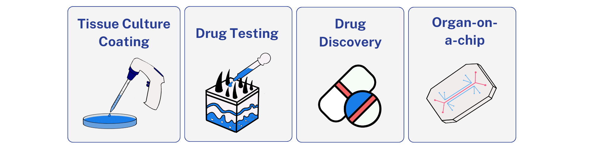
Applications:


")
Background
Product Description
Type I Collagen is the most abundant type of collagen in the human body, as a major structural matrix protein in skin, and many other tissues (bone, tendon, and fibrous connective tissues) [1]. There are a number of types of collagen identified to date, and all are composed of molecules containing three polypeptide chains arranged in a triple helical conformation [2]. The types of collagen differ slightly in the primary amino acid sequence of their polypeptide chains. Type I collagen is a heterotrimer composed of one α2 (I) chain and two α1 (I) chains [3].
Various types of cell surface receptors present on diverse types of mammalian cells recognize the collagen triple helix structure and facilitate cell attachment to collagen materials, including films and scaffolds. The most characterized cell receptors are the integrins α1 β 1 and α2 β 1 [4]. Many cell types express both forms of integrin, including mesenchymal stem cells (MSCs), fibroblasts, endothelial cells, chondrocytes, osteoblasts, and lymphocytes[3, 5-7]. Smooth muscle cells interact with collagen via α1 β1 , and epithelial cells attach via α2 β 1 [8, 9]. Collagen type I is characterized by the presence of three regions on SDS-PAGE corresponding to molecular weights. Alpha region has a molecular weight of 100 KDa and consists of two α1 chains and one α2 chain. Beta region has a molecular weight of 200 KDa and consists of two α1 chains fused together and α1 α2 chains fused together. Gamma region has a molecular weight of 300 KDa and consists of overlapping two α1 and one α2 chains. HumaCoat has all three regions and chains with very minimal fragmented proteins in between. HumaCoat is ideal for many applications including coating tissue culture surfaces to support cell attachment and growth or for making hydrogel.
Tissue Source
Type I collagen is isolated from human skin sourced strictly from American Association of Tissue Banks (AATB) accredited and FDA registered tissue banks and organ procurement organizations (OPOs). HumabiologicsTM strives to meet research needs by providing high quality biomaterials obtained from tissue partners who comply with requirements for transplantable human tissues under 21 CFR 1271 of the Food and Drug Administration (FDA).
Precautions and Disclaimer
HumaCoat is obtained from human tissue that has been tested and found negative for minimum of HIV-1 and -2, hepatitis B, and hepatitis C, as well as other infectious agents. Please review the Safety Data Sheet (SDS) for information regarding hazards and safe handling practices. HumaCoat is for research use only and is not intended for human use, diagnosis, screening, household, food or other uses.
Storage Conditions/Expiration
HumaCoat should be stored at 2-8 °C upon receipt. Do not freeze collagen solution. Research grade products have a shelf life of 6 months upon reciept.
- Bornstein, P. and H. Sage, Structurally distinct collagen types. Annu Rev Biochem, 1980. 49: p. 957-1003.
- Tanzer, M.L., Cross-linking of collagen. Science, 1973. 180(4086): p. 561-6.
- Heino, J., The collagen family members as cell adhesion proteins. Bioessays, 2007. 29(10): p. 1001-10.
- Barczyk, M., S. Carracedo, and D. Gullberg, Integrins. Cell Tissue Res, 2010. 339(1): p. 269-80.
- Heino, J., The collagen receptor integrins have distinct ligand recognition and signaling functions. Matrix Biol, 2000. 19(4): p. 319-23.
- Ivaska, J., et al., Cell adhesion to collagen. Is one collagen receptor different from another? Vol. 30. 1998. 273-283.
- Goessler, U.R., et al., In vitro analysis of integrin expression in stem cells from bone marrow and cord blood during chondrogenic differentiation. J Cell Mol Med, 2009. 13(6): p. 1175-84.
- Ebert, E.C. and A.I. Roberts, Human intestinal intraepithelial lymphocytes bind to mucosal mesenchymal cells through VLA4 and CD11A. Cell Immunol, 1996. 167(1): p. 108-14.
- Gilcrease, M.Z., Integrin signaling in epithelial cells. Cancer Lett, 2007. 247(1): p. 1-25.
Instructions for Use
Collagen-Coated Tissue Cultureware
Note: The following are general recommendations. Researcher should optimize parameters based on their specific applications. Optimal collagen concentrations for tissue culture surface coatings may depend on application and cell type and must be determined for each application. Typically, 10 – 100 µg/ml of collagen is used.
- Dilute HumaCoat with 10-20 mM HCl with a pH of 1.9-2.1 to 0.1 mg/ml. It is recommended to use ultrapure sterile water to make the 10-20 mM HCl.
- Mix by shaking or vortexing until the solution is homogeneous.
- Aseptically add collagen solution to tissue culture surface to evenly cover entire surface.
- Cover and incubate collagen coated surfaces at 37 °C for at least 2 hours. The optimal incubation time may vary depending on the experiment and cell type and should be determined for each application.
- Transfer collagen coated surfaces to biosafety cabinet and aspirate excess solution from coated surface careful not to scratch the surface. Remove lid to allow for air drying overnight.
- Rinse the coated culture surface with sterile PBS or culture medium to remove residual acid.
- Use coated cultureware immediately or keep sterile and store at 2-10 °C.
Quality Testing
| Test | Specification |
|---|---|
| Appearance (form) | |
| Color | Clear |
| Form | Solution |
| Solution Turbidity | Clear to Slightly Opaque |
| Concentration (mg/ml) | 1.0-1.5 |
| pH | 1.9-2.1 |
| Cell Attachment | Pass |
| Purity by SDS-PAGE & Coomassie Stain (alpha, beta and gamma regions with α1 & α2 chains) |
>95% |
| Sterility – USP 71 Modified Method | No Growth |
| Immunology/Serology Testing** | |
| HBV | Non-Reactive |
| HCV | Non-Reactive |
| HIV-1 | Non-Reactive |
| HIV-2 | Non-Reactive |
| RPR-Syphilis | Non-Reactive |
* Gel data is available upon request
** Testing was done using FDA approved kits in CLIA certified lab
*** Donor info is available upon request
Publications
- Patrawalla, N.Y., et.al. Funct. Biomater. 2023, 14, 363. https://doi.org/10.3390/jfb14070363
- Baltazar, T., Jiang, B., Moncayo, A., et al. Bioeng Transl Med. 2022;e10324. doi:10.1002/btm2.10324
- Baltazar, T., Kajave, N.S., Rodriguez, M., et al. J Biomed Mater Res. 2022; 110( 10): 2323-2337. doi:10.1002/jbm.b.35080
- Schmitt, T., Kajave, N., Cai H.H., Gu L., Albanna M., Kishore V. Journal of Biomaterials Applications. 2021;35(8):912-923. doi:10.1177/0885328220959162
- Ali, S., Patrawalla, N.Y., Kajave, N.S., et al. Biomacromolecules 2022 23 (12), 5137-5147 doi: 10.1021/acs.biomac.2c00985
Safety Data Sheet